Could T-wave morphology on a single-lead ECG heartbeat help to identify people at risk of sudden cardiac death?
A joint collaboration involving researchers from Queen Mary University of London, University College London (UCL) and the University of Oulu has identified T-wave morphological variations (TMV), measured from a single beat single-lead electrocardiogram (ECG), as a strong predictor of sudden cardiac death (SCD) in low- and high-risk populations.
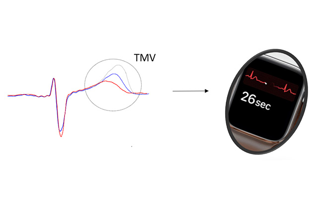
T-wave morphology variations on the ECG signal could enable large-scale screening to predict SCD risk using smartwatches.
Every year in the UK thousands of people die of sudden cardiac death (SCD), where the heart develops a chaotic rhythm that impairs its ability to pump blood around the body.
A new study, published in the Journal of the American Heart Association, used data from the UK Biobank, which includes health and genetic information from over half a million participants from across the UK, and from the ARTEMIS study, a cohort of ~2,000 Finnish patients with coronary artery disease. The researchers found that TMV is the only ECG marker measured at rest that associates with malignant ventricular arrhythmias in ~52,000 middle-aged individuals without heart disease from the UK Biobank. TMV was also a stronger SCD predictor in ~2,000 patients with coronary artery disease than traditionally used risk factors, like the interval or left ventricular ejection fraction.
First author Dr Julia Ramírez, from Queen Mary University of London, said: “These findings indicate a strong clinical translation potential of TMV, as its algorithm could easily be integrated into smartwatches and wearables for population screening.
“The impact of these results is important, because it enables a cheap and simple diagnostic method to early risk stratify individuals at SCD risk. We are now actively looking for additional cohorts of patients and different ancestries to validate them.”
The ECG signal reflects the electrical activity of the heart and T-wave morphology reflects the repolarization of the ventricles. A higher value of TMV indicates a larger electrical heterogeneity in the ventricles and a higher susceptibility to suffering from malignant ventricular arrhythmias and sudden cardiac death.
The study was conducted by the “Electrogenomics group” - a joint collaboration of investigators from Queen Mary University of London (Julia Ramírez, Patricia Munroe and Andrew Tinker) and UCL (Stefan van Duijvenboden, Michele Orini and Pier Lambiase), together with researchers from the University of Oulu (Antti Kiviniemi, Juhani Junttila, Juha Perkiomaki and Heikki Huikuri).
More information
Research publication: “ECG T-wave Morphology Variations Predict Ventricular Arrhythmic Risk in Low- and Moderate-risk Populations”. Ramírez J et al, Journal of the American Heart Association.